



The worst thing a doctor ever said to me about my back was “good news, your MRI is completely normal.” I was still in pain. I had driven there in pain, I would drive home in pain, and the scan that was supposed to explain it had instead, apparently, erased it, because a clean image seemed to mean the problem was in my head. It wasn’t. It took a second opinion and a longer conversation to learn what that first doctor should have said, which is that a normal scan is a real and useful piece of information that does not, on its own, mean nothing is wrong.
That gap, between what imaging shows and what pain actually is, is the whole subject here. Chronic pain conditions are genuinely difficult to diagnose because of their complex and varied causes, and body imaging is one of the most valuable tools medicine has for sorting them out. But it is a tool with a specific job and specific blind spots, and understanding both is what separates a patient who gets answers from one who gets dismissed. So this covers what imaging does brilliantly, and then the part almost nobody explains: why the scan can come back clean while the pain stays entirely real.
What Body Imaging Actually Does Well

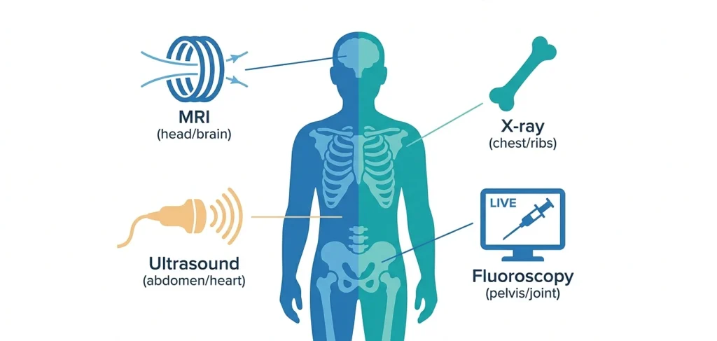
Imaging earns its central role in pain diagnosis by making the invisible visible, and each technique has a specific talent.
MRI is the detailed one. Using strong magnetic fields and radio waves rather than radiation, MRI produces detailed views of soft tissue, nerves, and joints, which makes it the go-to for spinal and nerve-related pain. It’s genuinely excellent at showing the physical health culprits that pain sometimes has: herniated discs pressing on nerve roots, spinal stenosis, joint degeneration, tumors, and fractures. When your pain follows a nerve down a leg or an arm in a clear pattern, MRI is often the tool that finds the compression causing it.
Other techniques cover what MRI doesn’t. X-rays read bone quickly and cheaply, which suits fractures and advanced joint changes. Ultrasound looks at soft tissue in real time, useful for tendon and muscle problems like tendinitis. And structural evaluation pairs with functional tests like nerve conduction studies to assess conditions such as carpal tunnel syndrome, where the question is less about a structure’s shape than about how well a nerve is actually working.
There’s also a treatment role that’s easy to overlook: imaging doesn’t just find problems, it guides the fix. Fluoroscopy provides live, moving X-ray during procedures, so when a physician places a corticosteroid injection or a nerve block, they can watch the needle reach the exact spot in real time, which makes the treatment both safer and more precise. That’s imaging working as a set of hands, not just eyes.
Why a Clean Scan Doesn’t Mean You’re Fine
Here’s the part my first doctor skipped, and it’s the more important half of the whole topic. Pain is produced by your nervous system, not by a photograph of your anatomy, and those two things come apart more often than most patients are ever told. A normal scan rules some things out. It does not rule out pain.
Start with what MRI physically cannot see. It images structure, the shape of things, at a certain resolution, which means it’s effectively blind to several real sources of pain: nerve excitability, inflammatory chemistry at the cellular level, and central nervous system sensitization all sit below or outside what the picture captures. A nerve can be chemically irritated and misfiring while looking structurally pristine. Early tissue changes and micro-tears can be too small to register. The image is honest about shape and silent about function, and pain is often a function problem.
Then there’s the phenomenon behind a lot of stubborn chronic pain central sensitization, where the nervous system itself becomes amplified, turning up the volume on pain signals so that the experience continues even after any original injury has healed or when no clear injury exists at all. This is the mechanism underlying conditions like fibromyalgia, and a standard structural scan of the painful area shows nothing, because the problem isn’t in the area, it’s in how the whole system is processing signals. In studies of chronic low back pain, a meaningful share of patients have entirely normal MRIs despite persistent, disabling symptoms, and researchers attribute those cases to exactly these non-structural causes: myofascial dysfunction, central sensitization, and other processes imaging wasn’t built to detect.
If your scan is clean and your pain is real, the honest next step is not another scan of the same spot. It’s a thorough clinical evaluation, a detailed history, a physical exam, sometimes a targeted diagnostic injection to test a specific structure, and consideration of central pain mechanisms. A normal image redirects the investigation. It does not close it.
The Trap That Runs the Other Direction

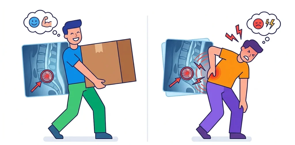
The disconnect cuts both ways, and this half is just as important to know, because it protects you from the opposite error: treating a scan finding as the automatic cause of your pain.
Imaging routinely finds “abnormalities” in people who feel completely fine. Disc herniations, bulges, and degeneration show up frequently on scans of people with no pain whatsoever, and studies of asymptomatic individuals find surprisingly high rates of exactly the findings that get blamed for pain in symptomatic ones. Thoracic disc herniations, for instance, appear in a substantial share of people with no symptoms at all. This means a finding on your scan is a suspect, not a verdict. A herniated disc visible on your MRI might be causing your pain, or it might be an innocent bystander your body has quietly ignored for years, and only clinical correlation, matching the image to your actual symptom pattern, can tell which. This is precisely why a good clinician treats the scan as one input weighed against the exam and your history, never as a standalone answer, and why two people with identical images can need completely different care.
What This Means for You as a Patient
Put the two halves together and imaging lands in its honest place essential, powerful, and incomplete on its own. It finds the structural causes it’s designed to find, guides treatments with real-time precision, and rules important things out. It also misses function, misses sensitization, and sometimes points at findings that aren’t the real problem. That’s not a failure of the technology. It’s the technology’s actual specification, and knowing it changes how you advocate for yourself.
A few things worth carrying into your own care:
- A normal scan is information, not dismissal. If you’re told your imaging is clean and offered nothing further while you’re still in pain, that’s the moment to ask about non-structural causes and clinical evaluation, not the moment to conclude you’re imagining it.
- A finding on a scan is a lead, not a conviction. Before accepting that a disc or a degeneration is your pain source, ask whether it actually matches your symptom pattern, because plenty of them don’t.
- The best diagnosis blends the picture with the person. Imaging works best alongside a clinical evaluation and other diagnostic tools, as part of a complete approach rather than the whole of it.
One closing note, offered plainly, since chronic pain is exhausting enough without being disbelieved: if you’re living with pain that your scans haven’t explained, that does not mean the pain isn’t real or that answers don’t exist. It means the answer may lie in the things imaging can’t photograph, and the right response is a fuller conversation with a healthcare provider, ideally one who treats a clean scan as the start of the investigation rather than the end of it. Talk to your provider about how body imaging fits your particular situation, and if a normal result ever gets used to wave you away, that is your cue to keep asking, not to stop.